Marcelo HURTADO, Laura JOUVIN, Ram Kumar PARI, Raphael DE ARAUJO BEAUVILAIN ALVES DE SOUZA and Roxane SYLVESTRE will receive a doctoral grant to conduct their PhD in CARe’s partner laboratories in Toulouse, in close cooperation with renowned laboratories abroad. We wish them every success in the realization of their PhD project.
PhD offer at the Toulouse Institute of Pharmacology and Structure Biology
The Ph.D. student will be enrolled in the CARe program.
Deadline for application: April 25th 2023
Starting date: September/October 2023
Working Place
The IPBS (Institute of Pharmacology and Structure Biology) is a joint research unit of the National Center for Scientific Research (CNRS), and the University of Toulouse. IPBS is located in Toulouse, a vibrant city in the south of France. The IPBS hosts about 250 scientific and administrative staff, including Ph.D. students and post-doctoral fellows of multiple nationalities. IPBS comprises 18 research teams, and the work carried out at the institute is dedicated to the discovery of new therapeutic targets in the fields of cancer, infectious diseases, and inflammatory diseases (https://www.ipbs.fr/). The selected Ph.D. candidate will work in the research team “Epigenetic Mechanisms in Cancer,” led by Priyanka Sharma at the IPBS, Toulouse. The candidate will be supported by a short training in the laboratory of molecular biophysics directed by X. Salvatella at the Biomedical Research Institute (IRB), Barcelona, Spain.
Technical methods
- Single-cell RNA transcriptomics.
- Live cell imaging and machine learning tools for analysis.
- Biophysical characterization of transcriptional condensates.
- Chromatin immunoprecipitation assays.
Expected Profile
Highly motivated and dynamic Ph.D. candidate with great communication skills and a strong interest in unraveling epigenetic mechanisms using multidisciplinary approaches.
Interested candidates, please contact at for further information.
Open position for a PhD in organic chemistry toward in vivo imaging
The Fundamental and Applied Heterochemistry Laboratory (LHFA), Toulouse, France and the Department of Drug Design and Pharmacology, University of Copenhagen, Denmark, are currently looking for candidates to enter a 3-year PhD program within the CARe Graduate School.
The successful candidate will be hired in a multicenter research program between Toulouse and Copenhagen bringing together chemists, radiochemists and biologists. The transdisciplinary research project targets the development and application of innovative chemical and biochemical tools for in vivo imaging of metabolic activity in the context of age-related diseases. The key idea is to combine easy-to-handle fluorescent or radioactive probes with biological vectors in a highly modular fashion. It will consist in the organic synthesis of both targeting and recognition units, as well as their bioconjugation in vitro or in vivo for further biological investigations.
The expected background of the successful candidate will be rooted in organic synthesis (connection to biological investigations would also be welcome). A sound knowledge of main group chemistry would also be beneficial. A strong ability to adapt to different research fields will also be of paramount importance. Although mainly based in Toulouse (France), the PhD training will partly take place in Copenhagen (at least 3 to 6 months).
Applications of students from all origins and of all genders nearly completing their master degree are strongly encouraged.
Deadline for submission: 28th of April, 2023.
Further information can be requested from:
A PhD position is open at Infinity to work on T-Follicular Regulatory cell heterogeneity in defective Ageing humoral response
The PhD student will be enrolled in the CARe program.
Deadline for application: April 15th 2023
Starting date: September/October 2023
Working context:
Infinity (Toulouse Institute for Infectious and Inflammatory Disease) is joint research unit of the French National Institute of Health and Medical Research (Inserm), the National Center for Scientific Research (CNRS), and the University of Toulouse. Infinity is a dynamic research institute comprised of 14 teams about 300 people and is located in Toulouse, a lively and fast-growing city in the Southwest of France with the 2nd largest student population. Within Infinity, the PhD candidate will work in the research team “Antigen Presenting Cells in T cell responses” led by Nicolas Fazilleau. This project is in close collaboration with ImmunXperts, a Q² Solutions company, localized at Gosselies (Belgium). During the PhD program, an internship of several months will be performed in this biotech company.
Working project:
The PhD candidate will study the development and biological roles of the human Tfr pool during physiological and ageing humoral responses.
Germinal centers (GCs) are the lifeblood of protective humoral immunity, require complex regulation that can decline with time, increasing vulnerability to infectious diseases and limiting vaccination benefits of elderly population. The integrity of GCs relied upon T follicular regulatory cells (Tfr), a regulatory subset that ensure antibody affinity for pathogens while decreasing self-recognition. Despite displaying a dual function in GCs reaction, Tfr cells were originally described as affiliated exclusively to Tregs (“natural” Tfr cells, nTfr), a population expressing autoreactive T-cell receptors (TCRs). More recently, we challenged this dogma by the description, in human tonsils, of a Tfr subset, descending from Tfh cells (“induced” Tfr cells, iTfr). However, the biologic role of a heterogenic Tfr pool in human and its impact on ageing immune responses are currently unknown.
The long-term goal of the proposed PhD program is to determine the biological role of the Tfr pool heterogeneity during GC response in humans and how ageing impacts this balance contributing thus to immune dysfunction. Our working hypothesis is that two distinct pathways shape the human Tfr pool, and immunopathology could result when one developmental pathway takes the lead. Our final ambition is to develop strategies to influence the Tfr pool composition that will improve the vaccine response of the elderly population.
Technical approaches:
– Single cell RNA sequencing on human T follicular populations
– Multiplex microscopy of human tissues
– Development of human lymphoid organoïds and in vitro functional assays
– Recruitment and immunophenotype by multiparametric flow cytometry of an « ageing” cohort
Expected profile:
We are looking for a highly motivated and dynamic PhD candidate with strong interest in immunology and excellent communication skills. Previous experienced with data analysis in R would be appreciated.
Interested, want to know more, how to apply
Contact us @
Congratulations to the two first CARe students to defend their thesis!
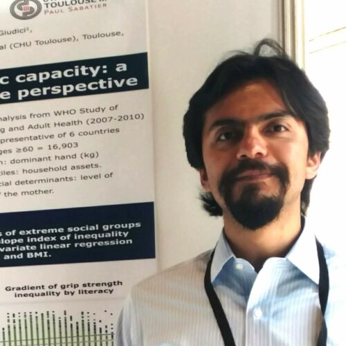
Emmanuel Gonzalez Bautista and Lea Da Costa Fernandes were the first two PhD students of the CARe Graduate School to defend their thesis.
Emmanuel’s thesis defense took place on December 5th, 2022, on the subject « Screening for abnormalities of intrinsic abilities with advancing age within the framework of the WHO ICOPE program: example of the locomotion approach », under the supervision of Pr. Sandrine Andrieu and Philipe de Souto Barreto, from the Centre for Epidemiology and Research in Population Health – CERPOP.
Lea defended her thesis on March 21st, 2023, on « Modeling the inflammatory response dynamics following tissue damage in adult mammals », under the supervision of Pr. Beatrice Cousin of the Restore Institute, and the co-supervision of François Peres, from the National School of Engineering in Tarbes – ENIT.
We congratulate them on this achievement and wish them every success in their future career.
Launch of CARe’s 2023 call for PhD proposals 2nd phase
The pre-selected projects in the framework of CARe’s 2023 call for PhD proposals are available here.
PhD candidates for the pre-selected projects will submit the full proposal to Claire Mendoza () and Clemence Grosnit () using this template. PhD candidates must be identified and are asked to send a complete CV including references.
At this step, PhD candidates must be identified and are asked to send a complete CV. Special attention will be given to candidates coming from foreign universities or countries. In addition, it is mandatory to provide a full description of the 3-6 months internship, including a signed attestation from the hosting foreign university or from industry.
The files must be submitted as a single PDF before May 12th, 2023. Applicants will defend their proposal in early June 2023, in front of a jury representative of the CARe Graduate school, with delegates from Toulouse partner doctoral schools. The audition will consist of 12 minutes of presentation and 15 minutes of questions. The presentation and the answers to questions will be done in English.
The presentation must include, in this order, 1 title slide, 1 slide presenting the candidate. The rest of the slides are dedicated to the presentation of the project (scientific justification of the research project, strategy, methodology, feasibility and risk management), including a description of the internship abroad or in industry. The following criteria will be evaluated by the jury: discussion of a previous research experience, quality of the oral presentation and of the response to questions.
Our 2023 call for PhD proposals is open
2023 Call for PhD proposals
The EUR CARe PhD program is devoted to the training of students in multidisciplinary research topics, from basic science to clinical or pharmacological applications, with a focus on cancer, ageing and/or rejuvenation. PhD research projects are directed by CARe-associated research teams from academic laboratories or partner companies. The EUR CARe PhD program selection will have two phases:
Phase 1: PhD pre-proposals will be submitted to Claire Mendoza () and Clemence Grosnit () using this template, before evaluation and selection by the CARe Scientific Committee. Deadline for application is February 24th, 2023. Upon acceptance, the proposers will be informed, and the proposals will be posted on-line on the EUR CARe website and social networks.
The applications must fall in the field of cancer, ageing and/or rejuvenation. In addition, multidisciplinary is mandatory and proposals must be at the interface of biology and mathematics, computer science, chemistry or physics. Examples are: chemists, physicists, computer scientists or mathematicians addressing complex biological questions by developing new molecules, tools, technologies or models to address biological questions, possibly doing some bench work, or biologists trained to advanced bioinformatics to develop new algorithms that would be later questioned/validated in experimental biological settings…
Partnerships between teams from different doctoral schools, foreign universities or industry are strongly encouraged (see scoring below). A short internship abroad (3 to 6-month) is mandatory, the nature of which should be briefly mentioned at this step. The travel and accommodation will be funded by the CARe program.
The scoring system (out of 10) is the following:
- Scientific quality of the proposal (6 points)
- Co-supervision (max 4 points) by teams from different doctoral schools (2 points), or with foreign university or industry (4 points).
NB: the candidates should not be formally identified at this step.
Phase 2: PhD candidates for the preselected projects will submit the full proposal to Claire Mendoza () and Clemence Grosnit () using the template that will be made available on CARe’s website.
At this step, PhD candidate must be identified and are asked to send a complete CV. In addition, it is mandatory to provide a full description of the 3-6 months internship, including a signed attestation from the hosting foreign university or from industry.
The files must be submitted as a single PDF before May 12th, 2023. Applicants will defend their proposal in early June 2023, in front of a jury representative of the CARe graduate school, with delegates from Toulouse partner doctoral schools.
The audition will consist of 12 minutes of presentation and 15 minutes of questions. The presentation and the answers to questions will be done in English.
The presentation must include, in this order, 1 title slide, 1 slide presenting the candidate. The rest of the slides are dedicated to the presentation of the project (scientific justification of the research project, strategy, methodology, feasibility and risk management), including a description of the internship abroad or in industry. The following criteria will be evaluated by the jury: discussion of a previous research experience, quality of the oral presentation and of the response to questions.
PhD fellowship at the interface between Cancerology / Immunology / Vascular biology M/F
3 years full time PhD student position funded by LABEX TOUCAN: Laboratoire d’Excellence Toulouse Cancer
starting date: October 1st, 2021
is available in the Teams of Jean-Philippe GIRARD: IPBS, CNRS and University of Toulouse, France and Camille LAURENT: CRCT, Inserm and University of Toulouse
Project Title: SINGLE CELL MAPPING OF THE VASCULATURE IN AGGRESSIVE B LYMPHOMAS AND TOPOLOGICAL INTERACTIONS WITH CD8 T CELLS AND y8 T CELLS
Keywords: aggressive B lymphoma, high endothelial venule, endothelial cell, spatial transcriptomics, scRNA-seq, anti-tumor immunity)
Abstract: Immunity plays an important role in cancer control, notably with highly mutated tumours such as Diffuse Large B-cell Lymphoma (DLBCL), an aggressive (fast-growing) non-Hodgkin lymphoma that affects B cells. Indeed, CD8 T cells and γδ T cells display potent antitumour cytotoxicity and are specifically reactive to lymphomas. DLBCL often develops in the lymph nodes. High endothelial venules (HEVs) are specialized blood vessels for lymphocyte entry into lymph nodes. HEVs may thus play an important role in the recruitment of CD8 T cells and γδ T lymphocytes in DLBCL lymph nodes.
The major objective of the project is to perform single cell mapping and spatial transcriptomics (ST) to characterize the density, maturation and functional status of HEV endothelial cells (MECA-79+CD31+) and non-HEV endothelial cells (MECA-79-CD31+), and define their topological interactions with cytotoxic γδ T cells and CD8 T cells in DLBCL patient’s biopsies. The project will greatly benefit from the expertise of:
– Jean-Philippe Girard’s team (IPBS) on HEV blood vessels, scRNASeq analyses of endothelial cells, and HEV-mediated lymphocyte recruitment in lymph nodes (Moussion and Girard, Nature 2011; Girard et al., Nat Rev Immunol 2012; Lafouresse et al., Blood 2015; Veerman et al., Cell Rep 2019), and
– Camille Laurent’s team (CRCT) on human B cell lymphomas (Laurent et al., Blood. 2020; Laurent et al., J Clin Oncol. 2017; Laurent et al., Blood. 2011), cytotoxic T cells, single cell RNA sequencing (scRNASeq, CITEseq) (Pizzolato et al., PNAS 2019; Pont et al., Nucleic Acids Res 2019) and spatial transcriptomics (transcriptomic hallmarks of all cells in a lymphoma biopsy in situ).
This study will deeply increase our knowledge about the distribution and functional status of HEV and non-HEV endothelial cells, γδ and other cytolytic T cells infiltrating human lymphomas. It may have important clinical consequences such as the identification of new predictive biomarkers, and guide T cell-based immunotherapies currently developed in Comprehensive Cancer Centers worldwide.
Requirement: Master in Cancerology, Immunology, Vascular Biology or Molecular Biology. Experience in scRNA-seq and/or bioinformatics will be a plus. We are looking for a creative and highly motivated PhD student strongly committed to research. Joint PhD supervisors: Drs Jean-Philippe Girard (IPBS) and Camille Laurent (CRCT).
Contract: 3 years full time PhD student position funded by LABEX TOUCAN (starting date: October 1st, 2021). The salary is in accordance with the French Ministry for Higher Education and Research salary scale. Social security and health benefits are included. Work context: research conducted at both IPBS and CRCT, two large Research Centers from CNRS, Inserm and University of Toulouse. All the necessary biological resources and research facilities, including state of the art technological facilities, will be available.
Deadline: the position will remain open until filled; only successful applicant will be contacted
How to apply: Please, send your application (in French or English, including a motivation letter, curriculum vitae, and rankings in University Licence/Master or Engineering school).
Multi-OMICs to unveil the role of lipid metabolism in the control of senescence and cellular plasticity in melanoma
PhD proposal
PhD supervisor : Nathalie ANDRIEU, CRCT: Toulouse Cancer Research Center, Toulouse, France
PhD co-supervisor : Gemma Fabrias, IQAC-CSIC: Institute for Advanced Chemistry of Catalonia, Barcelona, Spain
Visit us on
*
CARe PhD Proposals
How to explore physiological aging? A new framework for an in-depth explainability of machine learning models
PhD proposal
PhD supervisor : Chantal Soulé-Dupuy – IRIT, Toulouse
PhD co-supervisor : Paul Monsarrat – Restore, Toulouse
Aging is an overly complex biological process, involving multiple mechanisms at different levels, from molecular to tissue scale. With the increase of life expectancy, the challenge is to predict the physiological age to reach healthy aging, namely the one’s ability to adapt and efficiently respond to stressors. The recent use of machine learning (ML) strategies may accurately model the physiological age to prevent age-related disruption. However, black boxes properties of most ML algorithms do not support the understanding of their internal decision-making mechanisms (i.e., the explainability), crucial to highlight the critical variables on the prediction.
Using a strategy based on the coalition of attributes we recently develop algorithmic solution, whose performances and prediction qualities are superior to the gold standard SHAP (Shapley Additive exPlanation) approach. We propose to consider the prediction explanations, not as a final objective, but as a new data space allowing to impulse a better interaction between the ML model and the end user. This thesis project will focus on the development of an original framework for the biomedical community to explore in depth predictions of health evolution with age using an ML model. It will allow the detection of subpopulations with specific predictive explanations, organizing them hierarchically to ensure easy exploration. As a proof of concept, we will apply this framework to physiological age (in-house database of >40,000 subjects, 200 variables), to better understand biological determinants of ageing, their interactions, putative causal chains, and underlying physio-pathological mechanisms.
Key words: aging, machine learning
*
Visit us on
*
